Neuroscientists Map Specific Population of Brain Cells Governing Biological Sleep Pressure
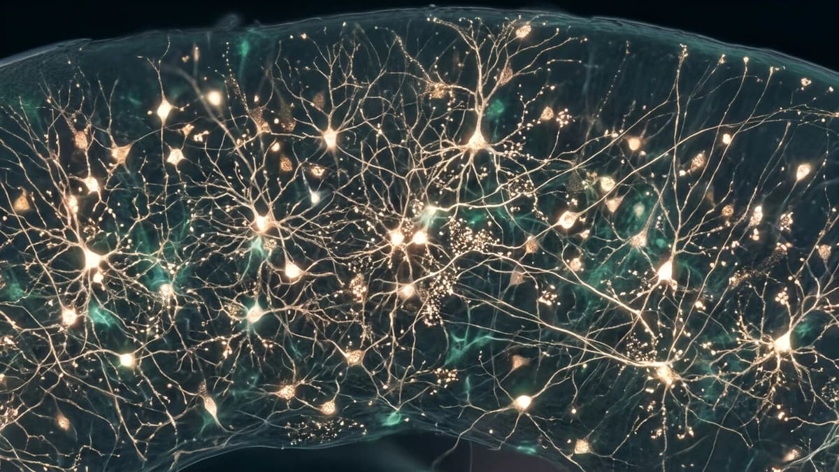
Neurobiologists at the University of Zurich and the Department of Neurobiology at Harvard Medical School identified in September 2026 a distinct subpopulation of deep-layer pyramidal neurons situated within layer 5b of the primary somatosensory cortex that functions as the brain's internal odometer measuring homeostatic sleep need in mammals. The discovery elucidates the precise biochemical cascade through which the mammalian brain measures the metabolic strain of prolonged wakefulness, demonstrating that these specialized cells synthesize, bind, and track interstitial adenosine while activating calcium/calmodulin-dependent protein kinase II (CaMKII). Once adenosine accumulation crosses a critical biochemical threshold following 14 to 16 hours of continuous wakefulness, this localized neuronal circuit emits synchronized low-frequency oscillations that inhibit ascending cholinergic and monoaminergic arousal nuclei within the brainstem, driving the transition into deep non-REM slow-wave sleep (N3).
The discovery resolves a longstanding debate in sleep physiology, challenging the traditional hypothesis that sleepiness results from an unstructured, diffuse exhaustion distributed uniformly across the entire cerebral cortex. In preclinical optogenetic validation trials, targeted photoactivation of this somatosensory circuit induced immediate slow-wave delta rhythms even in fully rested animal models, whereas selective chemical inhibition prolonged wakefulness without triggering the psychomotor impairment, tremor, or anxiety typical of conventional stimulant drugs.
The Enigma of Sleep Homeostasis and the Classical Two-Process Model
Ever since chronobiologist Alexander Borbély proposed the two-process model of sleep regulation during the early 1980s, medical science recognized that the sleep-wake continuum is governed by two interacting regulatory forces: Process C (the circadian rhythm) and Process S (the homeostatic sleep drive). While Process C has been thoroughly mapped to rhythmic transcriptional-translational feedback loops of Clock and Bmal1 genes within the hypothalamic suprachiasmatic nucleus under photic retinal synchronization, the precise anatomical seat and cellular mechanism generating Process S remained elusive.