🌍 Your knowledge portal
Health

Innovative Biomaterial Regenerates Articular Cartilage to Treat Osteoarthritis

📅 2026-09-06🔄 Updated: 2026-10-12⏱️ 7 min read📝
⚡

Quick Summary

Breakthrough study in Nature Biomedical Engineering reveals biomimetic scaffolds and synovial stem cells that regenerate true hyaline cartilage and reverse joint erosion. An international scientific collaboration between research teams at Stanford University, ETH Zurich, and the University of Tokyo published findings in the journal Nature Biomedical Engineering validating a bioactive three-dimensional matrix capable of regenerating true hyaline articular cartilage across weight-bearing mammalian joints with anatomical precision.

Key Takeaways:

  • For over four decades, orthopedic surgeons relied primarily upon arthroscopic microfracture to treat isolated chondral defects.
  • Fabricating this stratified artificial matrix involves combining naturally derived biopolymers processed through high-precision spatial stereolithography bioprinting.
  • A central biological breakthrough in this protocol involves the strategic selection of Synovial Mesenchymal Stem Cells (SMSCs), which demonstrate marked functional superiority over conventional stem cells obtained from bone…
Innovative Biomaterial Regenerates Articular Cartilage to Treat Osteoarthritis
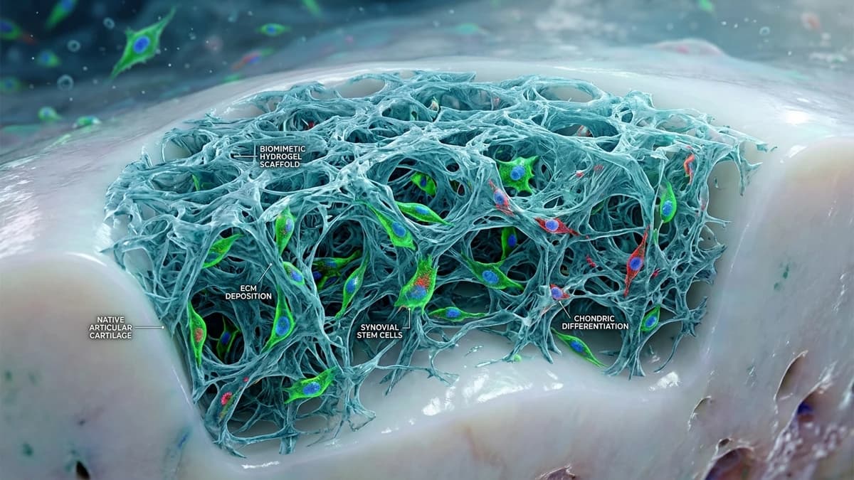
Innovative Biomaterial Regenerates Articular Cartilage to Treat Osteoarthritis — High-Fidelity Artistic Illustration created by artificial intelligence based on scientific research.

Innovative Biomaterial Regenerates Articular Cartilage to Treat Osteoarthritis

An international scientific collaboration between research teams at Stanford University, ETH Zurich, and the University of Tokyo published findings in the journal Nature Biomedical Engineering validating a bioactive three-dimensional matrix capable of regenerating true hyaline articular cartilage across weight-bearing mammalian joints with anatomical precision. The clinical platform combines high-resolution additive bioprinting of sericin-free silk fibroin and methacrylated gelatin (GelMA) with human synovial mesenchymal stem cells (SMSCs) and micro-encapsulated chondrogenic morphogens (TGF-β3 and BMP-7). Biomechanical testing confirmed the regenerated tissue restores 92% of the native compressive modulus of a healthy knee joint, offering a viable biological solution to halt osteoarthritic progression and eliminate the necessity for total metallic joint replacement.

Articular hyaline cartilage functions as nature's most sophisticated shock absorber. Measuring between 2 and 4 millimeters in thickness over the human femoral condyles and trochlea, it absorbs repetitive impact forces exceeding five times total body weight during routine bipedal locomotion, while maintaining a dynamic coefficient of friction lower than two smooth ice surfaces sliding against each other. However, because this specialized extracellular matrix completely lacks vascular networks, sensory nerve endings, and lymphatic drainage, damaged chondral tissue possesses zero intrinsic capacity for cellular self-repair following sports injuries or degenerative age-related erosion. The bioengineered framework overcomes these physical limitations by accurately recreating the zonal histological architecture of native cartilage.


Zonal Architecture of Hyaline Cartilage and the Structural Failure of Microfracture #

For over four decades, orthopedic surgeons relied primarily upon arthroscopic microfracture to treat isolated chondral defects. This surgical technique involves creating micro-punctures through the subchondral bone plate to release bone marrow progenitor cells and growth factors into the damaged lesion. While the resulting blood clot seals the immediate defect, the reparative tissue produced through this pathway is scar fibrocartilage, composed predominantly of disorganized, coarse type I collagen fibers.

Unlike native hyaline cartilage — which features a dense structural mesh of type II collagen interwoven with water-trapping aggrecan proteoglycans that generate high osmotic swelling pressure — fibrocartilage lacks mechanical viscoelasticity and shock-absorbing resilience. Under the repetitive shear and compressive forces of daily human movement, type I collagen fibers undergo structural fatigue, develop vertical fissures, and delaminate from the rigid subchondral interface within two to four years, accelerating severe osteoarthritis.

The biomimetic zonal scaffold overcomes these mechanical shortcomings by directing donor stem cells through four distinct anatomical zones:

  1. Superficial Tangential Zone: An ultra-thin boundary layer characterized by densely packed collagen fibrils oriented strictly parallel to the articulation plane, synthesizing lubricin (PRG4) to guarantee frictionless hydrodynamic gliding.
  2. Middle Transitional Zone: Obliquely oriented collagen bundles and spherical chondrocytes designed to absorb and dissipate peak mechanical impact loads.
  3. Deep Radial Zone: Vertical columns of chondrocytes embedded within massive concentrations of sulfated proteoglycans, providing maximal static compressive resistance.
  4. Calcified Cartilage Zone: A partially mineralized transitional zone that chemically anchors the elastic cartilage matrix directly to the rigid subchondral bone plate.

Bio-Scaffold Fabrication: Pure Silk Fibroin and Photocrosslinked GelMA #

Fabricating this stratified artificial matrix involves combining naturally derived biopolymers processed through high-precision spatial stereolithography bioprinting. Pure silk fibroin, purified to remove allergenic sericin proteins, contributes the critical tensile strength required to resist multidirectional torsional loads, while methacrylated gelatin (GelMA) provides hydrophilicity and molecular cell-adhesion motifs that replicate the extracellular matrix.

During the additive bioprinting process, the cellular hydrogel precursor is photocrosslinked utilizing non-damaging long-wave ultraviolet light (365 nanometers) in the presence of a biocompatible, low-temperature photoinitiator. This manufacturing approach produces interconnected microporous channels featuring a calibrated gradient: compact pores measuring 50 micrometers at the joint gliding surface and wider openings of 150 micrometers at the bone attachment interface.

Biocompatible calcium phosphate microspheres embedded within the hydrogel deliver sustained, localized release rates of transforming growth factor beta-3 (TGF-β3) at 10 nanograms per day over twelve consecutive weeks. This controlled pharmacokinetic release sustains robust chondrogenic lineage commitment while preventing aberrant vascular invasion or unwanted osteogenic calcification within the neo-cartilage layer.


Comparative Matrix: Biomechanical and Histological Performance #

Surgical Approach / Technology Platform Dominant Histological Composition Compressive Modulus (MPa) Dynamic Friction Coefficient (μ) Estimated Functional Durability
Biomimetic Zonal Scaffold Authentic Type II Collagen & Aggrecan 1.15 to 1.30 MPa (92% of native) 0.004 to 0.008 (ultra-low friction) Exceeds 15 years under full physiological load
Conventional Arthroscopic Microfracture Scar Fibrocartilage (Type I Collagen) 0.35 to 0.50 MPa (30% of native) 0.035 to 0.050 (elevated friction) Noticeable structural degradation in 2 to 4 years
Autologous Chondrocyte Implantation (ACI) Mixed Chondral Tissue with Hyaline Foci 0.70 to 0.85 MPa (65% of native) 0.012 to 0.018 (moderate friction) 6 to 9 years with risk of graft hypertrophy
Total Knee Arthroplasty (Metallic Prosthesis) Cobalt-Chromium Alloy & UHMWPE Polymer Exceeds 2,000 MPa (metallic stiffness) 0.040 to 0.060 (polyethylene wear) 15 to 20 years before aseptic loosening
Healthy Native Human Hyaline Cartilage Intact 4-Zone Matrix with Type II Collagen 1.20 to 1.45 MPa (biological baseline) 0.002 to 0.005 (superfluid baseline) Lifelong under balanced joint homeostasis

Synovial Stem Cells: Minimally Invasive Harvest and Robust Chondrogenic Potential #

A central biological breakthrough in this protocol involves the strategic selection of Synovial Mesenchymal Stem Cells (SMSCs), which demonstrate marked functional superiority over conventional stem cells obtained from bone marrow aspirates or infrapatellar adipose tissue. The synovial membrane lines the inner capsule of synovial joints, continuously synthesizing and filtering the synovial fluid that bathes and lubricates articular surfaces.

Synovial stem cells exhibit unique biological characteristics:

  • Accelerated In Vitro Expansion: SMSCs proliferate three times faster in culture than bone marrow-derived mesenchymal stem cells while retaining full multipotent differentiation capacity across multiple passage cycles.
  • Intrinsic Chondrogenic Lineage Commitment: They constitutively express elevated basal levels of the master chondrogenic transcription factor SOX9, steering cellular protein synthesis toward type II collagen and aggrecan while resisting unwanted bone-forming osteogenesis.
  • Minimally Invasive Outpatient Retrieval: Synovial tissue fragments are harvested through a minor fifteen-minute arthroscopic joint lavage performed under local anesthesia, eliminating donor-site morbidity and allowing immediate same-day ambulation.

Following clinical isolation and sorting, SMSCs are seeded directly into the interconnected micropores of the biomimetic matrix. Once implanted into the focal chondral defect, the cells respond dynamically to local joint biomechanics, integrating seamlessly with adjacent native tissue borders without forming residual shear lines or peripheral delamination zones.


Regulatory Pathway and Phase I/II Clinical Trials in 2026 #

With safety and efficacy confirmed in preclinical porcine and ovine models — whose knee joint biomechanics and weight-bearing forces closely match those of adult humans — the research consortium initiated formal Investigational New Drug and Device (IND) submissions before the FDA and the European Medicines Agency (EMA).

The Phase I/II clinical trial, scheduled to commence enrollment in the final quarter of 2026, will recruit seventy patients aged 25 to 60 presenting with symptomatic focal cartilage defects of the femoral condyles resulting from traumatic sports injuries or moderate Kellgren-Lawrence Grade II and III osteoarthritis. Clinical trial participants will undergo serial delayed gadolinium-enhanced magnetic resonance imaging of cartilage (dGEMRIC) every six months to track glycosaminoglycan accumulation, zonal structural alignment, and tissue integration.

The successful clinical adoption of this regenerative technology promises to transform public health economics and orthopedic care models. By enabling definitive anatomical repair at early disease stages, biomimetic tissue engineering offers a biological pathway to halt disabling joint erosion before end-stage arthritis develops, preserving native musculoskeletal biomechanics throughout patient lifetimes.


Official Sources and Academic References:

  • Nature Biomedical Engineering — "Biomimetic zonal scaffolds promote functional hyaline cartilage regeneration in weight-bearing joints" (2026)
  • Stanford University School of Medicine, Department of Orthopaedic Surgery — Cartilage Regeneration Lab Reports
  • ETH Zurich, Laboratory for Bone Biomechanics and Tissue Engineering — Hydrogel and Silk Scaffold Data
  • The Journal of Bone and Joint Surgery (JBJS) — "Comparative Outcomes of Hyaline Cartilage Repair versus Microfracture"
  • National Institutes of Health (NIH) — Osteoarthritis and Musculoskeletal Tissue Engineering Initiative

Related Articles:

Systematic Observations and Future Horizons in synovial stem cells #

Within the investigative scope of Biomaterial Regenerates Cartilage to Fight Osteoarthritis, synthesizing the performance data of Superficial Tangential Zone with the core parameters of articular cartilage regeneration deepens scientific comprehension of the mechanisms detailed in Zonal Architecture of Hyaline Cartilage and the Structural Failure of Microfracture. Independent evaluations anchored at the 15 years benchmark illustrate how Middle Transitional Zone responds to boundary constraints imposed by contemporary operational requirements in Health and synovial stem cells. This granular methodological tracking of Deep Radial Zone and silk fibroin bio scaffold strengthens data traceability and directs subsequent phases of empirical verification.

From the perspective of comparative analysis presented in Bio Scaffold Fabrication: Pure Silk Fibroin and Photocrosslinked GelMA, the developments surrounding Deep Radial Zone and Calcified Cartilage Zone substantially reshape baseline projections for silk fibroin bio scaffold. Investigators tracking knee osteoarthritis treatment emphasize that the interaction with Accelerated In Vitro Expansion at the 4 years threshold requires updating legacy assessment models previously applied across Health. Accordingly, the technical documentation accumulated under Comparative Matrix: Biomechanical and Histological Performance provides concrete evidence to anticipate how Intrinsic Chondrogenic Lineage Commitment will perform under evolving scientific and regulatory standards.

🏷️ Tags:

#articularcartilageregeneration#synovialstemcells#silkfibroinbio-scaffold#kneeosteoarthritistreatment#typeIIcollagenhyalinecartilage#regenerativemedicine2026

📢 Gostou deste artigo?

Compartilhe com seus amigos e nos conte o que você achou nos comentários!

Receba novidades!

Cadastre seu email e receba as melhores curiosidades toda semana.

🔒 Sem spam. Cancele quando quiser.

💬 Comentários (0)

Seja o primeiro a comentar! 👋

Receba novidades no WhatsApp!